While the vision chart tests your central vision, it does not provide information about the peripheral vision. The visual field test maps the sensitivity of the overall field of vision by projecting targets in your peripheral vision. Visual field test is most commonly performed for glaucoma but it is also valuable for assessing disorders of the retina, optic nerve and brain.
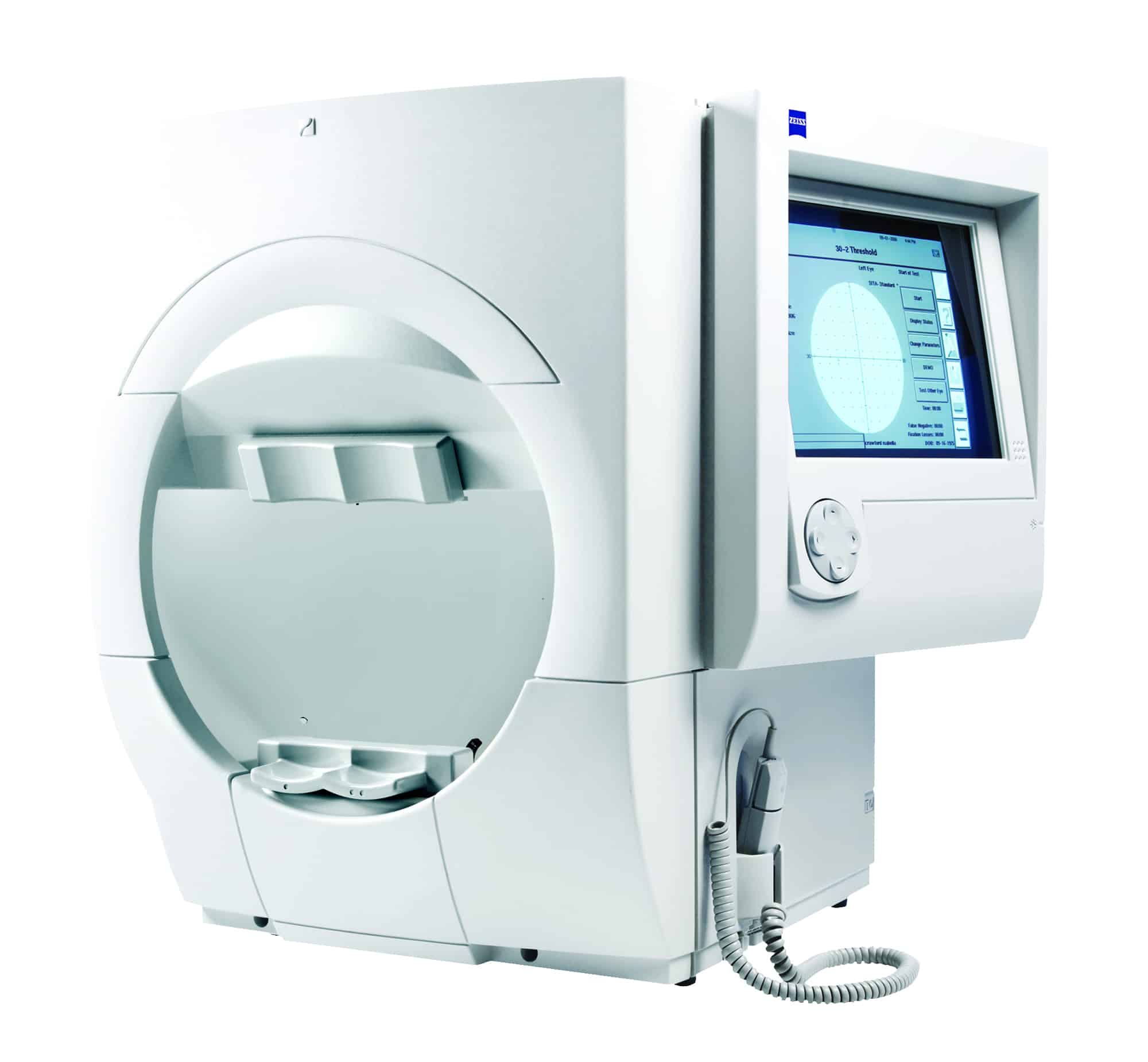
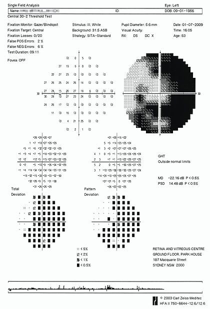
This specialised test maps the sensitivity of the entire field of vision by projecting small, controlled light targets into different areas, particularly in the peripheral (side) vision, while the patient focuses on a central point. The patient indicates when they see these targets, helping to create a detailed map of any vision loss or abnormalities.
The visual field test is most commonly used in the diagnosis and management of glaucoma, a condition that gradually affects peripheral vision and can lead to blindness if left untreated. However, it is also a valuable tool for detecting and monitoring other eye and neurological conditions. Diseases affecting the retina, such as retinitis pigmentosa, can cause characteristic patterns of vision loss that are detected through this test. Additionally, damage to the optic nerve from conditions like optic neuritis, tumours, or strokes can also lead to specific visual field defects, which can help in diagnosing underlying brain disorders.
By regularly performing visual field tests, ophthalmologists can monitor disease progression, adjust treatment plans, and ensure early intervention, when necessary, ultimately helping to preserve a patient’s vision and quality of life.