Retinal angiography is a safe and valuable tool that provides essential insights into vascular health, enabling early detection and effective management of vision-threatening conditions.
Angiography is an advanced diagnostic procedure used to evaluate the health of blood vessels and assess circulation throughout the body. In ophthalmology, retinal angiography plays a crucial role in detecting and monitoring various eye conditions that affect the blood supply to the retina and choroid.
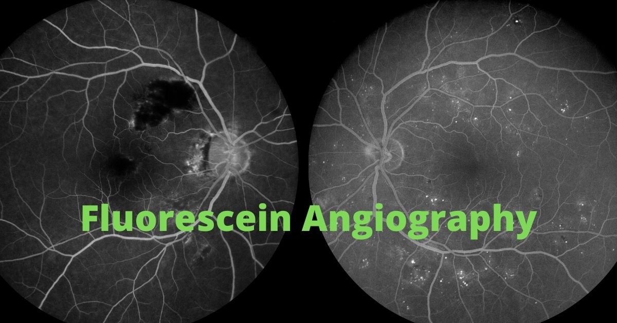
During the procedure, a special dye is injected into the bloodstream, allowing the blood vessels to become visible under specialized imaging. A high-resolution camera captures detailed images of the dye as it moves through the retinal and choroidal blood vessels, helping to identify abnormalities such as blockages, leaks, or abnormal growth of new blood vessels.
Fluorescein angiography (FA) is the most commonly used technique, where fluorescein dye highlights the retinal blood vessels, aiding in the diagnosis of conditions like diabetic retinopathy, macular degeneration, and retinal vein occlusion. In some cases, indocyanine green (ICG) angiography may be used instead, particularly when deeper layers of the eye, such as the choroidal circulation, need to be examined. This is especially useful in diagnosing conditions like central serous chorioretinopathy or certain types of macular degeneration.