Ophthalmic photography is an essential tool in modern eye care, allowing for precise documentation of eye diseases and their progression over time.
By capturing detailed images of the eye, this technique helps in diagnosing conditions, monitoring treatment effectiveness, and facilitating long-term patient care. The advancement of digital photography and computerized archiving has made this process more efficient, enabling eye specialists to track even subtle changes in a patient’s eye health with accuracy.
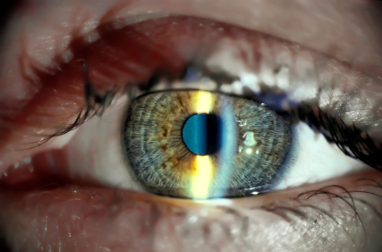
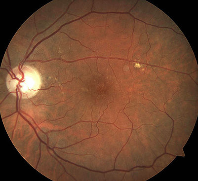
Depending on the reason for your visit, photographs may be taken of different parts of the eye. Images of the back of the eye, including the retina, macula, and optic nerve, are commonly used to assess conditions such as macular degeneration, diabetic retinopathy, and glaucoma. For certain conditions affecting the cornea, iris, or eyelids, high-resolution images of the front structures of the eye may be taken instead. These photographs serve as a valuable reference for both you and your doctor, helping to guide treatment decisions and ensure the best possible care for your vision.