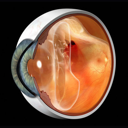
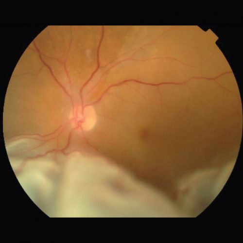
A retinal detachment occurs when the retina is pulled away from the inside wall of the eye. Left untreated, a retinal detachment will cause permanent and severe loss of vision.
Risk factors for retinal detachment?
- Short sightedness (myopia)
- Previous cataract surgery
- Severe eye injury or trauma
- Previous retinal detachment in your other eye
- Family history of retinal detachment
- Weak areas in your retina
- Warning symptoms of a retinal detachment – flashes and floaters


What are the warning signs of retinal detachment?

Sudden loss of vision or the appearance of flashes and floaters may be a sign of retinal detachment. Immediate eye examination is crucial.
What types of surgery do we use?
There are several ways to fix a retinal detachment, depending on the features of your condition.
There are 2 main surgical procedures undertaken to repair a retinal detachment:
Vitrectomy
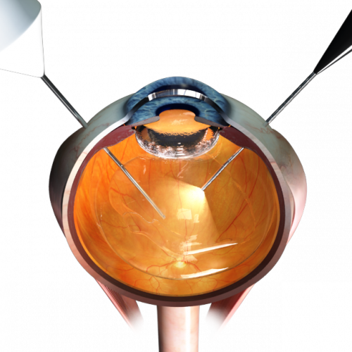
This technique is more commonly used and involves removing the vitreous using very fine instruments through wounds in the sclera (white of the eye). The retinal tears are identified and the fluid under the retina that caused the detachment are sucked out. A gas bubble is injected in the eye cavity to prevent fluid getting under the retina again. The gas bubble dissipates in 2 to 4 weeks and you may be asked to keep posturing (e.g. look down) for several days. Air travel is absolutely prohibited until gas is completely gone.

Scleral Buckle
This is an older technique but still is the most effective option for many types of retinal detachment. A piece of silicone rubber is sewn onto the sclera to push the wall of the eye inwards, bringing it closer to the detached retina. The buckle is permanently left in place. Buckle surgery is less comfortable than vitrectomy, and may lead to double vision.

